How Trauma Affects the Brain
When a client experiences trauma, it can seem like everything has changed – and to an extent, it has. . . on a neurobiological level. But that doesn’t mean there isn’t room to heal. So how can neurobiology inform trauma treatment?
Imagine, for a moment, that you are in session with a client.
They have a complex trauma history, including neglect and attachment trauma as a child.
As you’re wrapping up the session, you let the client know that you will have to reschedule next week’s appointment, as you will be out of town. But as you ask when else they might be available, you notice this client’s muscles begin to stiffen and their eyes go wide.
They have entered the freeze response.
As a clinician, you probably understand why this situation was triggering for the client – when their caregiver left as a child, they didn’t have their basic needs met for a long time – and so you might use your social engagement system to help the client back out of freeze.
But the client, on the other hand, may feel embarrassed, ashamed, or even confused about their response. So how can we help that client avoid a shame spiral?
The key just might be a little psychoeducation about the neurobiology of trauma.

Neurobiological Effects of Trauma
Trauma can become encoded in a client’s neurobiology, impacting their lives long after the threat has passed. But there are many different ways trauma can live in the brain – making it especially challenging to treat.
The Theory of the Triune Brain
Our understanding of the brain has changed considerably over the years – especially when it comes to the impact of trauma.
Theories are constantly being proposed and disproven. But even those that are now contested by many experts have still been useful stepping stones in our work to better understand the neurobiology of trauma.
One such theory is that of the triune brain. Paul MacLean, MD, proposed this concept, essentially simplifying the brain’s structures into three parts:
- Reptilian Brain : includes the brain stem and cerebellum.
The reptilian brain is responsible for survival and maintenance. It regulates the heartbeat, breathing, and other autonomic processes.
- Mammalian Brain :includes the subcortical region and limbic system.
The mammalian brain is responsible for emotion and memory formation. It allows us to learn.
- Primate/Human Brain : includes the neocortex.
The primate brain is responsible for executive and higher cognitive function. It regulates attention and focus, allows us to experience empathy , and enables complex reasoning and abstract thought.
So, let’s think back to the client who froze in session.
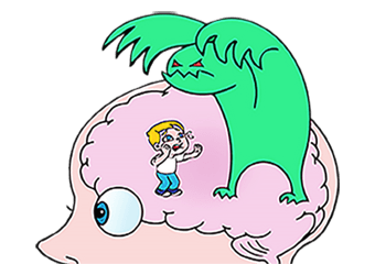
Their nervous system detected a threat, and streamlined that information to the reptilian brain, or “old brain,” as an immediate issue of survival. Then, their “old brain” automatically and unconsciously chose what seemed to be the most adaptive response in the moment: to freeze.
After the threat has passed, and we have re-established cues of safety in the office, the client’s primate brain, or “new brain” is back online, and they have the ability to reason, as well as to self-reflect.
And if their conscious mind disagrees with how their nervous system responded , or doesn’t understand why they ended up in freeze, that’s when our client might begin to feel shame.
Now, as we mentioned before, the triune brain model has received a lot of criticism[1] for being an overly simplistic generalization of a complex structure.
But while this theory may not be literal, the concept still stands. You see, there are certain structures in the brain that we share with reptiles, and those tend to be associated with our most primitive defense responses.
The Role of the Periaqueductal Gray
The periaqueductal gray is associated with the regulation of heart rate and blood pressure, autonomic processes like bladder control, pain reduction or inhibition, and – often incorporating many of these things – defense responses. This structure in the midbrain surrounds the part of the brainstem called the cerebral aqueduct.
As Ruth Lanius, MD, PhD, explains, “You have direct connections from the back part of the eye, the retina, to the brain stem. So, if you see something, even if you’re not aware of it, that can be transmitted to the brain stem immediately.”
Of course, the organism has to be prepared to defend itself in a fraction of a second. So even before we are consciously aware of the threat, the retina has already transmitted it to the superior colliculus, right next to the periaqueductal gray, which can mobilize fight-or-flight.
Bessel van der Kolk, MD, refers to this as “the cockroach part” of the brain, as it is our most primitive danger detection part. And when a client has been traumatized, the periaqueductal gray is almost always lit up. They are constantly in a state of defense.
The Brain Networks Trauma Tends to Target
According to Ruth Lanius, MD, PhD, trauma can damage three specific brain networks:
- Default Mode Network – the network which helps us process self-relevant information, like emotions or memories. If trauma has impacted this network, a client may not know what they are experiencing emotionally, feel disconnected from their body, or have trouble recalling memories. This can ultimately affect a client’s sense of self.
- Salience Network – the network which helps us figure out what in the environment is most important to respond to. Trauma often leaves this network overactive, so a client lives in a constant state of hyperarousal. This can impact a client’s relationships , as they may see threats where there are none. But in some cases, trauma can leave the salience network underactive as well, numbing them to cues of danger and leaving them more susceptible to future trauma.
- Central Executive Network – the network which helps us plan, think, concentrate, focus attention, and sustain short-term memories. With the impact of trauma, clients often have trouble sustaining attention – which is why childhood trauma is commonly misdiagnosed as ADHD. Clients may also have an increased tendency to dissociate.

How Trauma Impacts Telomeres
When clients experience trauma, it can impact every part of the mind and body – including telomeres.
As you know, our genes influence everything from our metabolism to what we look like. But each chromosome has a little structure, a telomere , helping it out.
Dan Siegel, MD, describes these structures like “the cap on a shoelace.”
It can be hard to imagine that such a small thing can have such a profound impact on both physical and mental health. But if you’ve ever had an old, worn pair of tennis shoes, you probably know what he means.
You see, with use, the little plastic cap at the end of the shoelace slowly deteriorates. And once the end of the lace is exposed, it begins to unravel.
Now with telomeres, the deterioration occurs as our cells replicate. Each new piece of the genome needs a protective cap, so they split and get shorter with each replication.
Eventually, our telomeres split to a point where they’re too short to protect the genes.
We have known for several years that adults with a history of PTSD tend to have shorter telomeres than those who haven’t experienced trauma. But a longitudinal study, led by Idan Shalev, PhD, further confirmed this phenomenon.
Children’s telomeres were studied at ages 5, 7, and 10. While there was visible shortening of all children’s telomeres as they aged, those who had reported exposure to violence had, on average, an increased rate of deterioration. In fact, those who were exposed to multiple types of violence had even shorter telomeres.
Researchers are still looking for ways to lengthen our telomeres. But in the meantime, there are plenty of other ways we can work with the brain to help clients heal from trauma.
The Dual Power of Neuroplasticity
Neuroplasticity is, essentially, the brain’s power to change.
As children, our brains are extremely malleable. And since we can’t speak, walk, or otherwise advocate for ourselves at birth, that ability for our brains to change is not just adaptive – it’s necessary for survival.

But neuroplasticity doesn’t disappear in the adult brain either. In fact, our brains are constantly changing in four distinct ways:
- Neurogenesis – certain regions of the brain continuously generate new neurons
- New Synapses – new experiences and skills can create new neural connections
- Strengthened Synapses – repetition and practice can strengthen neural connections
- Weakened Synapses – lack of use can weaken neural connections
Let’s think back to the beginning, with our example of the client who froze in session.
Unfortunately, the brain can’t tell the difference between the formation of good and bad habits. So as a child, every time they were in danger and the caregiver wasn’t there, the neural connection between those two phenomena was strengthened.
Many years later, as this client is in therapy working on rebuilding secure attachment , they begin to see their therapist as a caregiver. And the synapse – strong from its constant reinforcement in childhood – fires.
Then, if this client is ashamed of their freeze response, and repetitively thinks self-critical thoughts, the neural patterns for those self-critical thoughts will strengthen as well.
But as Norman Doidge, MD, says, “Plasticity is a gift of evolution.” Despite the so-called “dark side” of neuroplasticity, we can still create new connections. So, as we work with clients, we can think about it as creating and strengthening new synapses for healthier coping mechanisms.
How to Increase Neuroplasticity
Recent studies suggest that simple lifestyle changes may be able to increase the brain’s neuroplasticity.
Of course, neuroplasticity is an ever-changing field of research, and there’s a lot that we don’t know yet. But three strategies for increasing neuroplasticity look particularly promising:
-
Diet
Research suggests that certain foods may actually be helpful in fostering brain health and increasing neuroplasticity.
For example, a recent study[2] led by Daniela Ratto, PhD student, found that the progression of geriatric frailty in mice, both in terms of physical and cognitive function, could be slowed using the medicinal mushroom Hericium erinaceus, also known as the Lion’s Mane mushroom.
Furthermore, a series of studies by various researchers, including Paul Lapchak, PhD, et. al, have shown the spice turmeric to be beneficial for treating everything from insulin resistance in mice to increasing the lifespan and mobility of geriatric fruit flies. Of course, it isn’t turmeric itself that is so important, but rather the derived chemical, curcumin.

Now, these results have yet to be replicated in humans, but the prospects are rather hopeful.
-
Exercise
We often hear about exercise as a mood stabilizer. As much as we – and our clients – may resist it, we know it’s good for us. But not everyone realizes quite how much.
Bruce McEwen, PhD, has found extensive support for the benefits of exercise throughout his research. He even found that walking for 1 hour, 5 days a week can significantly change the size of the hippocampus, improving connections between short-term and long-term memory.
Furthermore, two separate studies, led by Thomas LaRocca, MD, PhD, and Christian Werner, PhD, have measured the long-term effects of exercise on telomere length.
Unfortunately, no matter how active young people were, their telomere length remained approximately the same. But when comparing results with middle-aged folks, the effects are staggering.
You see, those who did not exercise in middle age were found to have 40% shorter telomeres than young people, whereas those who regularly engaged in aerobic activity were only slightly shorter than their youthful counterparts.
Now, this doesn’t mean that exercise lengthens telomeres. It simply slows the rate at which they get shorter. Which, ultimately, still means that exercise can help protect our chromosomes from deterioration.
-
Mindfulness
As we know, trauma can leave clients with a constant sense of danger. They may become extremely anxious , or their defenses may be triggered by the smallest cue.
But, as it turns out, mindfulness[3] can actually help clients manage this activation in their brain.
Britta Hölzel, PhD, led a study in which clients with and without anxiety were exposed to images of facial expressions. Those with anxiety showed significantly higher amygdala activation than those without.
They saw ambiguity as a threat.
But after 8 weeks, those clients with anxiety who received a mindfulness-based stress reduction training showed significantly lower amygdala activation.
Research has shown how mindfulness practices tap into the brain’s natural neuroplasticity in a variety of other areas as well, including:
Neurofeedback
As it turns out, we can use operant conditioning on our own brains – with a therapy called neurofeedback.
If you think back to Psych 101, you may remember hearing about the Skinner Box experiment.
Basically, a young graduate student, B.F. Skinner, put a rat in a box. Inside, there was a lever which, when pressed, dispensed food. Of course, this experiment has been repeated in many variations over the years, but the results have been rather consistent.
Whether we’re using rewards or punishments to reinforce a specific behavior, operant conditioning is a rather effective training tool.
As Sebern Fisher, MA, has said, “It is more effective to reach the mind through training the brain than it is to reach the brain through training the mind.”
So how does neurofeedback therapy work?
When we say we’re conditioning the brain, we’re really focusing on the brainwaves.
As you likely know, there are several different kinds of brainwaves that we experience, and each is produced at a different frequency.
- Gamma: the highest frequency brainwaves, often associated with intense concentration.
- Beta: a high frequency brainwave, associated with anxiety or an otherwise active mind.
- Alpha: a mid-range frequency brainwave, often associated with relaxation and contentment.
- Theta: a low frequency brainwave, associated with deep relaxation and drowsiness.
- Delta: the lowest frequency brainwaves, indicating sleep.

Using an EEG, we can recognize the type of brainwaves a client is experiencing, and we can monitor changes.
Then, not unlike training a rat to press a lever, we reward the brainwaves at a certain frequency, often the alpha waves.
Of course, it’s also important to spend a portion of the session talking to the client, gauging their response to the last session, and adjusting accordingly.
At first, this experience will create a new synapse in the brain. But as the positive reinforcement continues, the synapse will be strengthened, encouraging the brain to create more of that kind of wave.
According to Sebern, this can take hundreds of sessions. She recommends seeing clients twice a week – but even that is nothing compared to the feedback receive every moment which has kept a client in a traumatized state so far.
Moving Forward with Brain-Based Trauma Treatment
There are many other strategies for using brain-based exercises to heal trauma, but ultimately, the brain still holds many mysteries.
Researchers are constantly finding new insights into the neurobiology of trauma – insights which will continue to prompt new strategies to help us better help clients heal.
And always remember – when you help someone heal from trauma, you can change the course of civilization. That’s because it’s not just that person’s life that changes, but that healing can also have an impact on their spouse, their children, and their friends and colleagues. And that can ripple out to their community, to their state and then to their country, and eventually to the world. What you do is so important.
References
Want more ideas and strategies you can use with your clients today?
To learn more about the latest strategies for treating depression, check out one of our courses:

The Neurobiology of Attachment
3 CE/CME Credits Available

Target the Limbic System to Reverse Trauma’s Imprint
3.25 CE/CME Credits Available

Expert Strategies for Working with Traumatic Memory”
2.75 CE/CME Credits Available

The Neurobiology of Trauma
3 CE/CME Credits Available